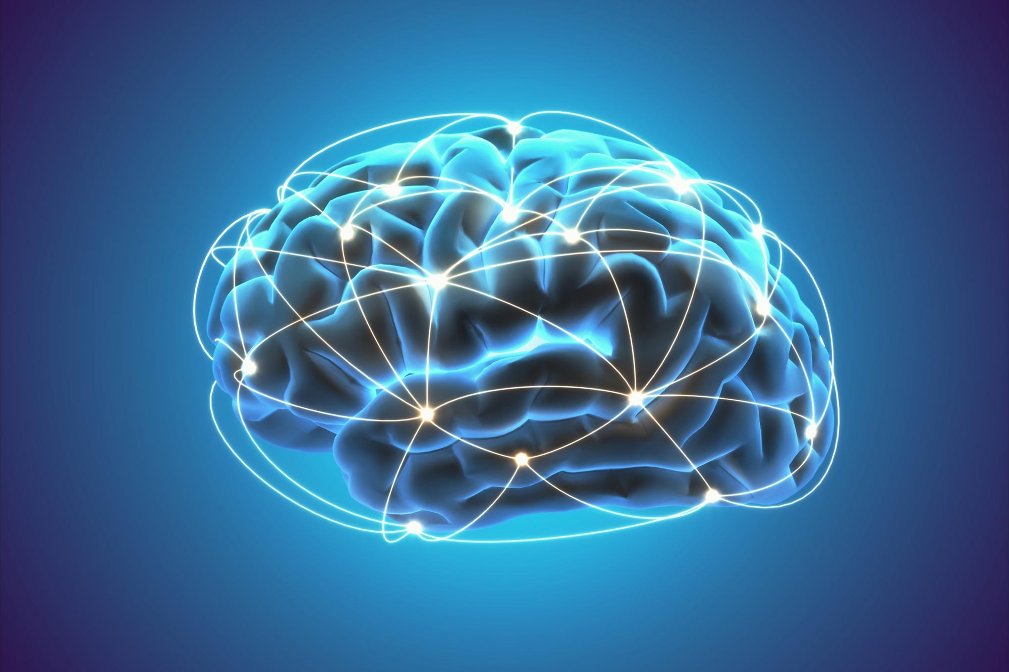
Human Brain Project (HBP) scientists used a unique multi-scale approach involving a variety of experimental methods to investigate the complex structure of brain connectivity. They combined techniques such as anatomical and diffuse magnetic resonance imaging, two-photon fluorescence microscopy, and 3D polarized light imaging to visualize and understand nerve fibers at different spatial scales. Their findings, incorporated into the Jülich Brain Atlas for reference, reveal new insights into the connectivity and function of different brain regions.
Researchers in the Human Brain Project used a multi-scale approach, from the molecular to the macro level, combining different imaging modalities to examine the connectivity, or interconnected structure, of the human brain. They used 3D Polarized Light Imaging to visualize nerve fibers and spatially map the data to the Jülich Brain Atlas, revealing new insights into brain structure and function.
Understanding how our brain works doesn’t involve examining how different brain regions are interconnected by nerve fibers. In the journal ScienceHuman Brain Project (HBP) researchers review the current state of the field, provide insights into how brain connectivity is structured at different spatial scales – from the molecular and cellular to the macro level – and evaluate existing methods and future needs. Understanding the complex system of connectivity.
„It’s not enough to study brain connectivity just once or twice,” Forschungszentrum Jülich et al. says Katrin Amunts, lead author and HBP Scientific Director of the Institute of Neurology and Medicine (INM-1) at the &O. Vogt Institute for Brain Research at the University Hospital Düsseldorf. „Connectivity exists at multiple levels. To understand its structure, we need to look at multiple spatial scales simultaneously by combining different experimental methods in a multi-scale approach and integrating the data obtained into multi-level atlases such as the Jülich brain atlas we created.
Markus Axer from Forschungszentrum Jülich and first author at the Department of Physics, University of Wuppertal. Science Essayn, with his team at INM-1, developed a unique method called 3D Polarized Light Imaging (3D-PLI) to visualize nerve fibers at microscopic resolution. With the aim of creating a 3D fiber atlas of the entire human brain, the researchers traced three-dimensional courses of fibers in serial brain sections.

Detail of a human brain section showing the architecture of single axonal fibers in the hippocampus revealed by 3D polarized light imaging. Colors represent 3D fiber orientations, highlighting the trajectories of individual fibers and trajectories. Credit: Markus Axer and Katrin Amunts, INM-1, Forschungszentrum Jülich
Together with other HBP researchers from Neurospin in France and the University of Florence in Italy, Axer and his team recently imaged the same tissue block from the human hippocampus using different methods: anatomical and diffusion magnetic resonance imaging (amRI and dMRI), two. -[{” attribute=””>photon fluorescence microscopy (TPFM) and 3D-PLI, respectively.
Microscopy methods like TPFM provide sub-micrometer resolution images of small brain volumes revealing microstructures of the brain’s cerebral cortex, but they have their limitations in disentangling fibers connecting distant brain regions, which build the deep white matter structures. This is even more true for electron microscopic measurements, which enable nanometre-resolved insights into a cubic millimeter of brain tissue. In contrast, dMRI can be used for tractography at the whole-brain level – visualizing white matter connections – but cannot resolve individual fibers or small tracts.
“3D-PLI serves as a bridge between micro and macro methods,” says Amunts. “This is because 3D-PLI resolves the fiber architecture at high resolution and, at the same time, allows imaging of whole-brain sections that we can then reconstruct in 3D to trace fiber connections.”
Combining dMRI, TPFM, and 3D-PLI enabled the researchers to superimpose the three modalities within the same reference space. “This integration of data was only made possible by imaging one and the same tissue sample,” explains Axer. The human hippocampus block traveled from Germany to France, back to Germany, and finally to Italy, being processed and imaged in different laboratories benefiting from the local, highly specialized equipment.
The researchers then used the Julich Brain Atlas to spatially anchor their data in an anatomical reference space. The three-dimensional atlas contains more than 250 cytoarchitectonic maps of brain areas and forms the centerpiece of the HBP’s Multilevel Human Brain Atlas. “Our brain atlas enables us to pinpoint exactly where in the brain we find these microstructures,” explains Amunts. The dataset is openly accessible via the HBP’s EBRAINS infrastructure and can be browsed in an interactive atlas viewer.
The researchers multi-scale approach combining multiple modalities at different spatial scales to unravel the human connectome is unique and provides exciting new insights into how the human brain works.
Even though the hippocampus reconstruction is a lighthouse project, there are several international efforts ongoing (or about to start) that need to be orchestrated at an open atlas level to enable the integration of multi-scale data. Amunts and Axer emphasize that this is a prerequisite for revealing the principles of connectivity within the experimentally accessible range of scales – from axons to pathways. In other words, an integrated multi-scale approach that combines micro and macro methods is necessary to describe and understand the nested organization of the human brain. This requires critical reassessment of current methodology, including tractography, the authors say.
Reference: “Scale matters: The nested human connectome” by Markus Axer and Katrin Amunts, 3 November 2022, Science.
DOI: 10.1126/science.abq2599

„Oddany rozwiązywacz problemów. Przyjazny hipsterom praktykant bekonu. Miłośnik kawy. Nieuleczalny introwertyk. Student.