Researchers at the University of Illinois Urbana-Champaign have introduced an AI technique that significantly improves the atomic force microscope (AFM), enabling it to visualize material features smaller than the tip of the probe. This breakthrough, which provides the first truly three-dimensional profiles beyond conventional resolution limits, promises to revolutionize nanoelectronics development and material exploration.
Atomic force microscopy, or AFM, is a widely used technique that quantitatively maps material surfaces in three dimensions. However, the precision of AFM is limited by the probe size of the microscope. A new artificial intelligence technique has been developed to overcome this barrier to enable microscopes to achieve higher resolution in material analysis.
A deep learning algorithm developed by researchers at the University of Illinois Urbana-Champaign was trained to remove the effects of probe width from AFM microscope images. As reported in the press nano letters, The algorithm outperforms other methods in providing the first truly three-dimensional surface profiles at resolutions below the width of the microscope probe tip.
Breakthroughs in Material Surface Imaging
„Accurate surface height profiles are important for the development of nanoelectronics and scientific studies of material and biological systems, and AFM is a key technique that can measure the profiles non-invasively,” said I. Professor of Materials Science & Engineering and U. Project Lead. „We've demonstrated how to be more precise and see even smaller things, and we've shown how AI can be used to overcome a seemingly insurmountable limit.”
Often, microscopy techniques can only provide two-dimensional images, essentially providing researchers with aerial photographs of object surfaces. AFM provides full topographic maps that accurately show the height profiles of surface features. These three-dimensional images are obtained by moving a probe over the object's surface and measuring its vertical deflection.
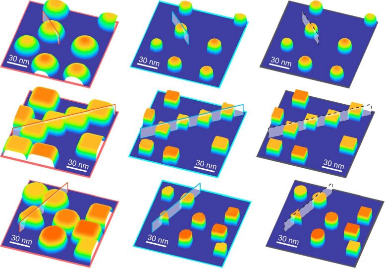
AFM images processed by a deep learning algorithm. The left column contains the simulated AFM images, the center column contains the images processed and reconstructed by the algorithm, and the right column contains the original images before the AFM effects are added. Credit: NanoLead. 2024, 24, 8, 2589–2595
If the surface features approach the size of the tip of the probe – about 10 nanometers – then they cannot be resolved by the microscope because the probe becomes too large to „feel” the features. Microscopists have known about this limitation for decades, but I. Researchers at the U.
„We turned to AI and deep learning because we wanted to get the height — the precision rigor — without the inherent limitations of more conventional mathematical methods,” said Lalit Bonagiri, a graduate student in Zhang's group and lead author of the study.
Deep learning algorithm
Researchers have developed a deep learning algorithm with an encoder-decoder architecture. It first “codes” raw AFM images by decomposing them into abstract features. After the feature representation has been manipulated to remove undesirable effects, it is again „decoded”.
A recognizable image.
To train the algorithm, the researchers created synthetic images of three-dimensional structures and simulated their AFM readings. An algorithm was constructed to transform simulated AFM images with probe size effects and extract fundamental features.
„We had to do something really buggy to achieve this,” Bonagiri said. „The first step in typical AI image processing is to rescale the brightness and contrast of images against some standard to simplify comparisons. In our case, absolute brightness and contrast were the meaningful part, so we had to drop that first step. It made the problem more challenging.
To test their method, the researchers synthesized gold and palladium nanoparticles with known dimensions on a silicon host. The algorithm successfully removed the probe tip effects and correctly identified the three-dimensional features of the nanoparticles.
„We've provided a proof of concept and shown how to use AI to significantly improve AFM imaging, but this work is just the beginning,” Zhang said. „Like all AI algorithms, it can be improved by training on better data, but the path forward is clear.”
Reference: Lalit Krishna Samant Bonagiri, Jirui Wang, Shan Zhou and Yingjie Zhang, 22 January 2024, “Nanoscale Precision Surface Profiling Enabled by Deep Learning” Nano letters.
DOI: 10.1021/acs.nanolett.3c04712
Carl R. The experiments were performed at the Wuss Institute for Genomic Biology and Materials Research Laboratory.
Supported by the National Science Foundation and the Arnold and Mabel Beckman Foundation.

„Oddany rozwiązywacz problemów. Przyjazny hipsterom praktykant bekonu. Miłośnik kawy. Nieuleczalny introwertyk. Student.