Nanomaterials such as Two-dimensional crystals and biomolecules share atomic-scale electrical polarization properties capable of producing significant macroscopic effects. These effects affect a variety of applications, from molecular biology and neuroscience to advanced photonic and energy storage devices.
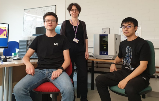
Dr Laura Fumagalli of the University of Manchester's Condensed Matter Physics Group is credited with developing the scanning electron microscope. This technique is used to study the dielectric properties of nanoscale materials based on atomic force microscopy (AFM) measurements of electrostatic force and current.
In 2021, Dr. Fumagalli needed a state-of-the-art AFM to increase his team's measurement capabilities. His primary requirements are atomic resolution in all three dimensions, method flexibility and sample environment control, including the ability to operate seamlessly in liq.
Image credit: Asylum Research – Oxford Instruments Company
Research methods
Dr. Fumagalli has extensively used Electromagnetic Force Microscopy (EFM) in his research to study the dielectric properties of various materials under the influence of voltage bias. For example, he used EFM to investigate interfacial and confined water.1 Through this research, Dr. Fumagalli and his colleagues have conclusively demonstrated for the first time that water near a surface is significantly less polar than bulk water.
This discovery has important implications in many fields, including biology and energy storage technology. Low-noise electronics in AFM are critical to maximizing signal-to-noise in data. Dr. Fumagalli used EFM to investigate the dielectric properties of biological molecules, including protein complexes and DNA.2
As he is affiliated with the Graphene Group and the National Graphene Institute in Manchester, Dr. Fumagalli is remarkably active in the field of 2D nanomaterials. He recently investigated 2D heterostructures, including 2D twisted layers, which have novel electro-optical properties and potential applications in the development of next-generation devices.3,4
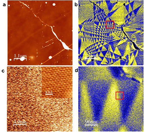
Throughout this research, he and his team used both EFM and Kelvin Force Probe Microscopy (KPFM) to visualize piezoelectric and ferroelectric properties, specifically the ferroelectric triangular domains in 2D twisted crystals as illustrated below.
Examples of images taken with Cyber ES on a twisted bilayer of MoS2 By Laura and her team. Using electrostatic force microscopy (EFM), they observed ferroelectric triangular domains (b,d), missing in the topographic image (a). Lateral force AFM images in (c) visualize the atomic structure of the layers—Zoom into the square area indicated in (d). Image credit: Asylum Research – Oxford Instruments Company
User registrations
To Dr. Fumagalli, The Cyber ES Environmental AFM proved to be an exemplary option in his laboratory. „First, it certainly brings the high resolution we need in our work. And importantly, it's reasonably easy to get this resolution on a routine basis without requiring special skills or touch—even our less experienced students can take publication-quality data without much training,” says Laura.
„Additionally, Cypher supports all the different methods we want, from simple morphology under air to more challenging nanoelectrical measurements in different ambient conditions—such as submerged samples. We really appreciate that the operating software is open source. I'm looking forward to improving and extending the AFM methods to interrogate the samples in more detail. Always looking, so using customized code is invaluable.
Notes and further reading
- L. Fumagalli et al. (2018) Anomalous Low Dielectric Constant of Confined Water, Science 3601339–1342. https://www.science.org/doi/10.1126/science.aat4191
- A. Cuervo et al. (2014) Direct measurement of dielectric polarization properties of DNA, PNASE3624–E3630. https://doi.org/10.1073/pnas.1405702111
- P ares et al. Piezoelectricity in single-layer hexagonal boron nitride Adv. Matter. 32 (1), 1905504. https://onlinelibrary.wiley.com/doi/full/10.1002/adma.201905504
- CR Woods et al. (2021) Charge-polarized interfacial superlattices in partially twisted hexagonal boron nitride, Nature Commun., 12, 347. https://doi.org/10.1038/s41467-020-20667-2
This information was obtained, reviewed, and modified from materials provided by Asylum Research, an Oxford Instruments company.
For more information about this source, please visit Asylum Research – An Oxford Instruments Company.

„Oddany rozwiązywacz problemów. Przyjazny hipsterom praktykant bekonu. Miłośnik kawy. Nieuleczalny introwertyk. Student.


